
Abdominal Ultrasound Emergency Room Imaging | Fort Worth ER
Abdominal ultrasound has become one of the fastest diagnostic tools in emergency medicine, helping ER physicians identify life-threatening conditions in minutes rather than hours. If you're experiencing sudden abdominal pain in Fort Worth or the surrounding communities, understanding how this imaging technology works can help you make informed decisions about your care.
In This Article
- What Is Abdominal Ultrasound in Emergency Medicine?
- Acute Abdominal Imaging: Conditions Emergency Ultrasound Detects
- Trauma Imaging and Point-of-Care Ultrasound Assessment
- How Abdominal Ultrasound Works in the ER Setting
- When to Seek Emergency Abdominal Ultrasound Evaluation
- Abdominal Ultrasound at ER of Fort Worth
- Frequently Asked Questions About Emergency Abdominal Imaging
- Frequently Asked Questions
⚡ Key Takeaways
- Abdominal ultrasound provides real-time imaging without radiation, making it ideal for point-of-care emergency assessment.
- Acute abdominal imaging can detect fluid collections, organ enlargement, appendicitis, gallstones, and other serious conditions within minutes.
- Trauma imaging via ultrasound helps ER physicians rapidly assess internal bleeding and organ damage after falls or accidents.
- 24/7 availability of emergency ultrasound at ER of Fort Worth means faster diagnosis and treatment decisions for abdominal emergencies.
- If you have severe, sudden abdominal pain, call 911 or go to the nearest emergency room immediately.
What Is Abdominal Ultrasound in Emergency Medicine?
Abdominal ultrasound is a safe, non-invasive imaging technique that uses high-frequency sound waves to create real-time images of organs, tissues, and fluid inside the abdomen. Unlike X-rays or CT scans, ultrasound produces no radiation, making it an ideal first-line diagnostic tool in emergency settings where speed and safety are equally critical.
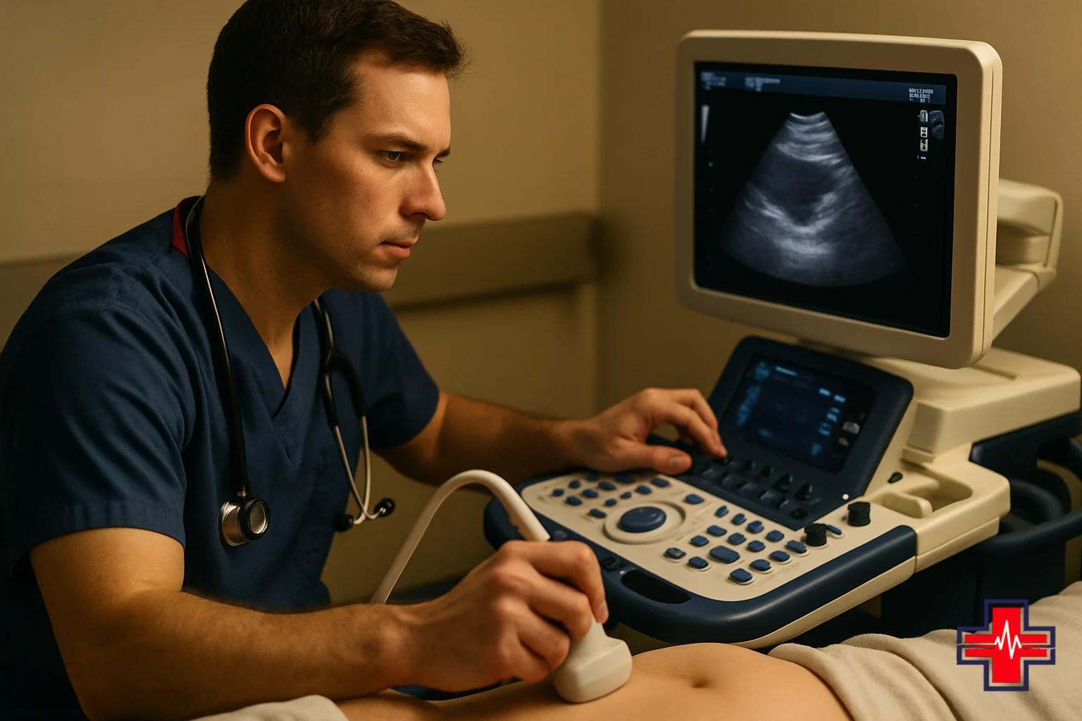
In emergency medicine, abdominal ultrasound is often called point-of-care ultrasound (POCUS) because the examination is performed by the emergency physician or ultrasound technician directly at the patient's bedside. This eliminates delays associated with transporting patients to a separate radiology department, allowing clinicians to make treatment decisions immediately based on visual findings.
The technique works by transmitting sound waves through the skin via a handheld transducer. These waves bounce off internal structures and return as echoes, which are converted into two-dimensional images displayed on a monitor. Emergency medicine specialists are trained to recognize normal anatomy and spot abnormalities that suggest serious, treatable conditions.
Acute Abdominal Imaging: Conditions Emergency Ultrasound Detects
Emergency physicians rely on acute abdominal imaging to rapidly identify life-threatening conditions that require immediate intervention. Abdominal ultrasound is particularly sensitive for detecting several critical presentations:
- Free fluid in the abdomen: Fluid collections (ascites) or blood accumulation suggest internal bleeding, organ rupture, or peritonitis and demand urgent evaluation or surgery.
- Appendicitis: An inflamed appendix appears as a dilated, non-compressible tube. Early detection prevents perforation and peritoneal infection.
- Cholelithiasis and cholecystitis: Gallstones visible within the gallbladder, or gallbladder wall thickening and pericholecystic fluid, indicate acute biliary disease causing severe pain.
- Abdominal aortic aneurysm (AAA): Ultrasound can measure the aortic diameter and detect aneurysmal dilation, a time-critical emergency.
- Ovarian pathology: Ovarian cysts, torsion, or rupture cause acute pelvic pain; ultrasound differentiates gynecologic emergencies from other abdominal causes.
- Urinary retention and bladder distension: Inability to void becomes evident on ultrasound, guiding catheterization decisions.
- Hydronephrosis: Kidney swelling from ureteral obstruction (kidney stone) is visible, confirming obstruction as the pain source.
Because these conditions can escalate rapidly, rapid and accurate acute abdominal imaging is critical to patient outcomes. Many emergency departments now include bedside ultrasound as part of their standard trauma and abdominal pain protocols.
Trauma Imaging and Point-of-Care Ultrasound Assessment
In trauma patients, every second counts. Point-of-care ultrasound has revolutionized the initial assessment of injured patients by detecting life-threatening internal bleeding without moving the patient off the stretcher or delaying care. This is especially important for trauma imaging in blunt injury cases.
The most common trauma ultrasound protocol is the FAST exam (Focused Assessment with Sonography for Trauma), which examines four key regions:
- Perihepatic view (right upper quadrant): Checks for free fluid around the liver, indicating internal bleeding from liver or right kidney injury.
- Perisplenic view (left upper quadrant): Assesses for fluid around the spleen, common in splenic lacerations.
- Pelvic view: Evaluates the pelvis for fluid suggesting massive internal bleeding.
- Pericardial view: Looks for fluid around the heart that could compress it, a sign of cardiac tamponade.
Trauma imaging via ultrasound takes only 2–3 minutes and is often performed while the trauma team is assessing airway and breathing. If free fluid is detected, it signals the need for immediate surgical intervention or transfer to a trauma center. For fall victims, motor vehicle accident patients, and others with blunt trauma, point-of-care ultrasound has become the standard of care because it is rapid, repeatable, and requires no patient transport.
How Abdominal Ultrasound Works in the ER Setting
The process of obtaining an abdominal ultrasound in the emergency room is straightforward and patient-friendly. After you check in and describe your symptoms, the ER physician may order an ultrasound as part of your diagnostic workup. No special preparation is usually needed in an acute setting, though fasting may improve image quality for gallbladder studies if time permits.
A trained ultrasound technician or ER physician will apply a clear gel to your skin to improve sound wave transmission. The transducer is then gently moved across your abdomen, and real-time images appear on the monitor. You may be asked to take a deep breath, change position, or point to areas of tenderness to help guide the examination. Most abdominal ultrasounds take 10–15 minutes, though focused emergency protocols can be completed in minutes.
Once imaging is complete, the ER physician reviews the images and discusses findings with you. If the ultrasound identifies a specific diagnosis—such as appendicitis, cholelithiasis, or free fluid from trauma—your treatment plan is adjusted accordingly. Results are available immediately, eliminating the hours-long wait typical of hospital radiology departments.
Because abdominal ultrasound uses real-time imaging rather than static snapshots, the emergency physician can watch organs move, assess blood flow with Doppler ultrasound, and measure structures dynamically. This interactive capability gives emergency medicine specialists powerful diagnostic insight in minutes.

When to Seek Emergency Abdominal Ultrasound Evaluation
If you are experiencing a life-threatening emergency, call 911 or go to the nearest emergency room immediately.
Sudden, severe abdominal pain always warrants emergency evaluation. Several red flags suggest you should seek immediate care at an ER rather than waiting for an appointment with your primary physician:
- Severe, sudden pain: Pain that comes on abruptly and is intense suggests acute pathology such as appendicitis, perforation, or obstruction.
- Pain with fever: Fever combined with abdominal pain points to infection (appendicitis, diverticulitis, peritonitis) that can deteriorate rapidly without treatment.
- Pain with vomiting or inability to keep food down: Persistent vomiting suggests obstruction or severe inflammation and risks dehydration.
- Pain after trauma or falls: Blunt abdominal injury can cause internal bleeding that develops over hours; early imaging detects life-threatening bleeding before shock occurs.
- Abdominal pain with severe back pain: Sudden, tearing abdominal and back pain radiating to the flank suggests aortic aneurysm, an immediate surgical emergency.
- Pain with signs of shock: Dizziness, rapid heartbeat, pale skin, or altered mental status with abdominal pain indicate serious bleeding or sepsis requiring emergency intervention.
- Pain in pregnant patients: Abdominal pain in pregnancy requires rapid evaluation to rule out ectopic pregnancy, placental abruption, or preterm labor.
The emergency physicians and ultrasound specialists at ER of Fort Worth are available 24/7 to evaluate acute abdominal pain. Whether you live in Fort Worth, Keller, Alliance, Saginaw, Haslet, Watauga, Roanoke, North Richland Hills, Haltom City, Bedford, Southlake, Grapevine, or Flower Mound, emergency abdominal imaging is available without appointment.
Abdominal Ultrasound at ER of Fort Worth
ER of Fort Worth, located at 4561 Heritage Trace Parkway, Suite 117, Fort Worth, TX 76244, offers immediate abdominal ultrasound and point-of-care emergency imaging 24 hours a day, 7 days a week. Our ER physicians and ultrasound technicians are trained in emergency medicine protocols and can perform focused trauma imaging, acute abdominal assessment, and comprehensive ultrasound examinations without delay.
At our facility, you can use our online check-in feature to streamline your arrival and reduce wait times. Once you arrive, our clinical team rapidly triages your condition and orders imaging based on your symptoms and physical examination. Because ultrasound is performed at the bedside, you don't have to leave the treatment area for imaging, and our physicians can integrate ultrasound findings into your care plan immediately.
Our experienced ER physicians understand that time is critical when you're in pain. By combining abdominal ultrasound with other diagnostic tools such as blood tests, physical examination, and clinical judgment, we rapidly identify the cause of your abdominal pain and begin appropriate treatment.
If you think you need emergency evaluation, call us at 817-945-4200 or come directly to our emergency room. We're here to help residents of Fort Worth and surrounding areas access world-class emergency diagnostic imaging when it matters most.
Frequently Asked Questions About Emergency Abdominal Imaging
Below are answers to common questions patients ask about abdominal ultrasound in emergency medicine settings.
Frequently Asked Questions
Is abdominal ultrasound safe?
Yes. Abdominal ultrasound uses sound waves, not radiation, and has no known harmful effects. It is safe for all patients, including pregnant women, making it an ideal first-line imaging tool in emergency settings. Unlike CT scans, there is no radiation exposure or risk of contrast allergies.
How long does an emergency abdominal ultrasound take?
A focused emergency ultrasound (such as a FAST exam for trauma) takes 2–3 minutes. A more comprehensive abdominal ultrasound evaluating organs and fluid takes 10–15 minutes. Results are available immediately, allowing the ER physician to begin treatment without waiting for radiology reports.
Can abdominal ultrasound detect appendicitis?
Yes. Ultrasound can visualize an inflamed appendix as a dilated, fluid-filled, non-compressible tube. Combined with clinical findings, ultrasound has high sensitivity and specificity for appendicitis, allowing rapid diagnosis and surgical referral when needed.
What if my ultrasound is normal but I still have pain?
A normal ultrasound is reassuring and often rules out serious abdominal pathology. However, some conditions (such as early appendicitis, early gastroenteritis, or musculoskeletal pain) may not be visible on ultrasound. Your ER physician will consider your symptoms, exam findings, and any additional tests to determine if further imaging or monitoring is needed.
Do I need to fast before an emergency abdominal ultrasound?
No. In emergency settings, ultrasound is performed immediately without fasting. For elective studies of the gallbladder, fasting helps image quality, but in acute situations, diagnosis and treatment cannot be delayed.
Is emergency ultrasound available at ER of Fort Worth on nights and weekends?
Yes. ER of Fort Worth operates 24/7, and abdominal ultrasound is available any time of day or night. You can walk in without an appointment or call 817-945-4200 if you have questions about your symptoms.